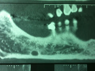
Maschio, 39 anni. Presenza di area radiotrasparente a margini netti nel contesto del corpo mandibolare, al livello del 2° - 3° molare.Reperto radiologico occasionale, nessuna sintomatologia:
RX Ortopantomografia
La lesione, indagata con TC mandibolare con ricostruzione dental scan, mostra di essere in diretta comunicazione con la loggia sottomandibolare.
Diagnosi: Lacuna di Stafne della mandibola
Terapia: in questi casi nessuna terapia è richiesta, tuttavia è corretto effettuare un follow-up radiologico annuale, poiché altre lesioni osteolitiche potrebbero mimare una lacuna di Stafne.
Stafne defect
Radiographically, it is a well-circumscribed radiolucency with a sclerotic border, and presents without any symptoms. It is usually a developmental defect of the jaw.The Stafne defect, first described in 1942,[1] is a depression of the mandible on the side nearest the tongue. It was previously known by many names, including static bone cyst,[2] Stafne idiopathic bone cavity,[3] and salivary gland inclusions in the mandible,[4] but is now known as a pseudocyst. The depression usually allows for the presence of a salivary gland. An early case of Stafne's defect has been discovered in a 7th century BC adult male individual from Klazomenai, one of the 12 cities of the Ionian League (now in modern Turkey) [5]]
References
^ Stafne, EC. Bone cavities situated near the angle of the mandible. JADA 1942;29:1969–1972.
^ Rushton, MA. Solitary bone cysts in the mandible. Br Dent J 1946;81:37-4
^ Barakat, N; AbouChedid, J. Cavite idiopathic mandibulaires. Rev Dent Liban 1973;23:35-40
^ Seward, GR. Salivary gland inclusions in the mandible. Br Dent J 1960;108:321-325


Questo commento è stato eliminato dall'autore.
RispondiElimina